[Special Feature]
Regarding tumor lesions, which are considered common urological diseases, atypical cases and rare diseases that require differential diagnosis may be encountered. This special feature provides practical information for today's practice, summarizing key points and precautions for differential diagnosis, as well as useful additional tests for these diseases!
Introduction by Yoshihiko Fukukura and Masahiro Jinzaki
Excerpt from the introduction: Tumor lesions in urological diseases can present atypical imaging findings due to background factors and pathological diversity, often making diagnosis difficult.
In this special feature, we cover a wide range of atypical cases, in addition to typical cases, and provide detailed explanations of noteworthy imaging findings and useful additional tests.
Renal Cell Carcinoma ● Daiyu Akita, Masahiro Jinzaki
Angiomyolipoma ● Ryo Yamamoto, Yoshihiko Fukukura, et al.
Renal Pelvis and Ureteral Cancer ● Yukiko Honda, Shogo Maeda, et al.
Bladder Cancer ● Hiroshi Shigesato
Prostate Cancer ● Hiromi Edo
Adrenal Cortical Adenoma and Pheochromocytoma ● Kota Yokoyama, Ukihide Tateishi
Testicular Germ Cell Tumor ● Kaori Yamada
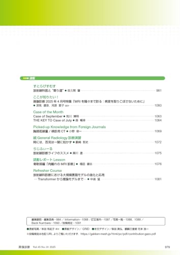
[Series]
Strabismus
A "Detour" with a Radiologist ● Hajime Sakuma
Here's What You Need to Know!
April 2025 Issue of Diagnostic Imaging: "Examining Every Corner of MRI: Avoid Missing Lesions"
● Tatsuya Fukami, Aiko Kawahara, et al.
Case of the Month
Case of September ● Hiroaki Arakawa
THE KEY TO Case of July ● Nobuyuki Mori
Picked-up Knowledge from Foreign Journals
Low-Dose Chest / Screening CT ● Shuichi Ono
Continued General Radiology Diagnostic Exercises
Sometimes, seeing is believing ● Tetsushi Yabusaki
Radifile
Recommendations for a Radiological Diagnostic Life ● Ryo Kurokawa
Radiogram Report Lesson
Bone and Soft Tissue Edition: "MRI Diagnosis of Strained Muscles" ● Takeshi Fukuda
Refresher Course
Evolution and Applications of Large-Scale Language Models in Diagnostic Radiology
─From Transformers to Inference Models─ ● Takeshi Nakaura


![Images! - Expert Imaging Diagnosis In Obstetrics and Gynecology - March 2021 Issue [Magazine]: Obstetrics and Gynecology Special Edition](https://img.joomcdn.net/e02e6fddceeec440a37af440dd95008d4411e908_69_100.jpeg)